Dr. Ov Slayden’s Seminar 4/3/24
Professor, Oregon National Primate Research Center
Imaging and ablation of endometriosis in nonhuman primate models
Researchers in Oregon seek novel therapies for women’s most under-diagnosed disorder
Endometriosis is a disorder that affects roughly 10-20% of women during their lives. It is characterized by endometrial tissue (the innermost lining of the uterus that is shed during menstruation) adhering to tissues outside of the uterus, resulting in characteristic “chocolate-like cysts”. The symptoms of this disorder include heavy bleeding, ectopic lesion development, pain, and infertility. The most common theory is that endometriosis develops from retrograde menstruation (back flow) of endometrial fragments through the fallopian tubes. Endometriotic disease is difficult to diagnose because there are no reliable blood tests on the market. The most common diagnostic option throughout the world is laparoscopic surgery, and invasive technique where the surgeon views the lesions with a light and camera in the body cavity. As a result, diagnosis of the disorder is often delayed by more than 12 years after the onset of the disease.
Nonhuman primates (NHPs) are valuable models for human female reproductive research. Their large female reproductive organs (e.g. ovaries, uterus, and cervix) closely resemble human reproductive tracts, and unlike mice, both NHP and women menstruate and experience a similar menstrual cycle. These similarities make NHPs a promising model for endometriosis research. Spontaneous endometriosis is observed in Old-World monkeys including baboons and macaques. Using this information, Dr. Ov Slayden, a Professor at the Oregon National Primate Research Center, aimed to create a viable and relevant model of endometriosis with the end goal for improving diagnostic and therapeutic technologies for this historically under-diagnosed disease.
Using a technique called menstrual seeding, Dr. Slayden grafts endometrial tissue into menstruating rhesus macaques and observes roughly 50% of the NHPs develop these characteristic cysts. Using this technique, Dr. Slayden and his team of researchers explore potential diagnostic and therapeutic techniques. Using drug-carrying nanoparticles (<100 nanometers), the researchers can target endometriotic cell markers (e.g. a protein called KDR). Endometriotic patients show higher levels of KDR than those with non-pathologic menstruation, so nanoparticle-loaded with proteins can be used as a potential diagnostic target. Using these targeted nanoparticles with fluorescent payloads, a technician could visualize the lesion without the need for any surgical procedures.
The team also uses techniques such as infrared or magnetic ablation (resulting in specific tissue heating and destruction) to non-surgically remove the lesions. Used together, these nanoparticles have the potential for a non-surgical alternative for diagnostic, lesion ablation, and treatment of reoccurrence. In addition to the work already done in this model by this ambitious team, there are sure to be many new and innovative endometriosis-related research due to the development of this NHP endometriosis model. (written by Erin Carulli)
Dr. Shamsun Nahar’s Seminar 3/20/24
Post-doctoral Scholar, Drs. Jaewook Jeong and Taehoon Kim’s laboratories, OB/GYN & Women’s Health, University of Missouri
Tumor suppressor role of MIG-6 and oncogenic effect of b-catenin activation in endometrial cancer
MIG6: The secret molecule controlling endometrial cancer progression
Endometrial cancer arises when cancerous cells grow within the endometrium (the innermost layer of the uterus). Ranked as the 6th most prevalent cancer among biological women, with 67,000 new cases in 2024 so far, endometrial cancer is a disease that many individuals will one day face. There are four stages of endometrial cancer, the least invasive being stage 1, where the cancer cells are contained within the uterus, to stage 4, where the cancer has spread throughout the lymph and to secondary organs (e.g. the bladder, rectum, or lung).
Dr. Nahar, a post-doctoral scholar in the Department of Obstetrics, Gynecology and Women’s Health, University of Missouri, is investigating the molecular mechanisms that cause endometrial cancer, with the goal of identifying new methods to combat this disease. PTEN, a gene responsible for regulating tumor growth, is one of the most commonly mutated tumor suppressor genes, being found mutated in 60-80% of cancer cases. Additionally, MIG6 is a gene that is commonly found mutated in endometrial cancer. Looking at these genes, Dr. Nahar investigates their direct role in causing endometrial cancer.
She found that when MIG6 and PTEN are absent (called knockout) in mouse uterus, the mice had reduced survivability rates and accelerated tumor growth when compared to mice who also lost PTEN expression alone (the control group). Conversely, when MIG6 was overexpressed in PTEN knockout mice, the lab found that tumor progression was suppressed when compared to the control, suggesting that MIG6 controls the tumor progression and can prevent tumor growth. In addition to these two proteins, Dr. Nahar is also investigating roles of another protein called b-catenin in endometrial cancer. Ultimately, the work done by Dr. Nahar illuminated molecular mechanisms underlying endometrial cancer. This important research adds to the work being conducted by countless other dedicated scientists and will help the entire scientific community better understand how cancer occurs, how it spreads, and how we can treat it (written by Erin Carulli).
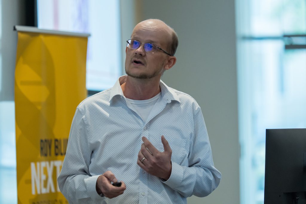
Dr. Michael Soares’s Seminar 3/6/24
Distinguished Professor, Institute for Reproductive and Developmental Sciences and Department of Pathology and Laboratory medicine, University of Kansas Medical Center
Trophoblast cells at the uterine-placental interface
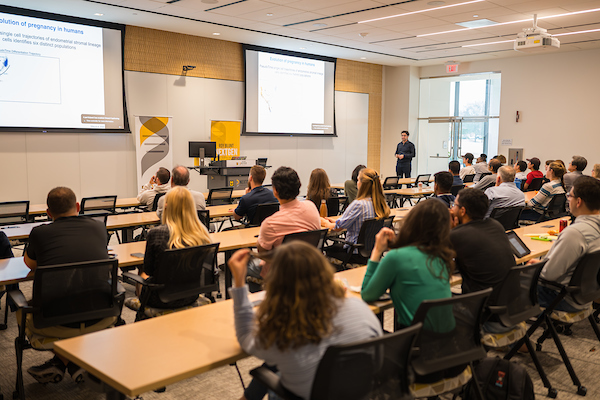
Invaders from within: how cells in the placenta breach the maternal defense (lay audience summary of the seminar below)
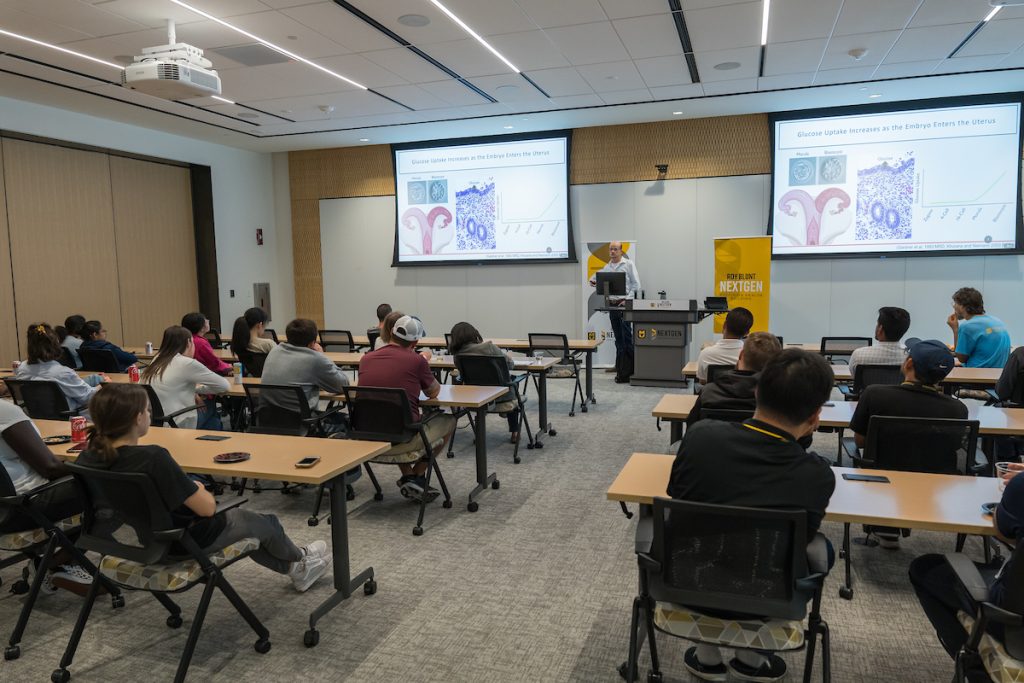
Trophoblast cells, which form in the outer layer of a blastocyst (the early stages of an embryo), are an important population of cells supporting pregnancy. Trophoblast cells go on to develop into a sizable portion of the placenta and are the first cells to differentiate within the fertilized egg. The hemochorial placenta, an essential barrier in maternal-fetal nutrient delivery, is subject to trophoblast invasion. Though these cells play necessary roles in embryo development, the mechanisms that regulate their function are still a mystery.
Dr. Michael Soares, Distinguished Professor, Pathology and Laboratory Medicine, University of Kansas Medical Center, aims to understand if there is a conservation at the uterine placental interface between rats and humans. This understanding will help identify if rats are a viable model to research trophoblast invasion in the hemochorial placenta, a process that is not present in all animals. Utilizing single-cell RNA sequencing, Dr. Soares found that both humans and rats experience invasive trophoblast cell populations at the hemochorial placenta.
With the conserved process in both rats and humans, trophoblast invasion in the hemochorial placenta can be studied using multiple genetic models in rats, such as gene ablation, to provide a better understanding of placental cell functions and behaviors in humans. With more understanding, we can find novel medical interventions to increase positive pregnancy outcomes or preventative methods for pregnancy loss.
Dr. MD Saldur Rahman’s Seminar 2/21/24
Assistant Research Professor, OB/GYN & Women’s Health, University of Missouri – Columbia
Therapeutic effect of nanceria as a non-steroidal anti-inflammatory drug (NSAID) on endometriosis in a mouse model
Nanoparticles are a girl’s best friend: improving diagnosis and treatment of endometriosis (lay audience summary of the seminar below)
Endometriosis is a chronic disease that affects one in ten biological women around the world. Normally, shed endometrial tissue (the innermost layer of the uterus) is expelled from the body during menstruation, but with endometriosis, the tissue remains in the body’s cavity and leads to lesions, pain, and infertility.
There are signaling molecules that can be targeted and depleted (knocked out) which have been shown to alleviate the symptoms of endometriosis (i.e. pSTAT3), but this type of intervention has not helped solve endometriosis-related infertility in mice. To address this shortcoming, Dr. Saidur Rahman, an Assistant Research Professor in the OB/GYN & Women’s Health Department, University of Missouri, has looked to nanoparticles to help identify and treat all of the symptoms of endometriosis, infertility included.
One advantage of using nanoparticles is the ability to visualize endometrial lesions without invasive surgery. Traditionally, endometriosis is diagnosed by symptoms with no definitive test to confirm the disease, which has led to many women missing diagnosis. To confirm endometriosis, doctors have had to perform surgery to view the lesions inside the body’s cavity. Using nanoparticles that can be injected and then travel specifically to endometrial lesions allows us to view the lesion without the need for surgery.
In addition to imaging, nanoparticles can deliver therapeutic payloads to endometrial lesions, mitigating off-target effects (such as infertility). Dr. Rahman found that injecting a nanoparticle called Nanoceria (a molecule which can reduce inflammatory effects specifically in endometrial tissue) was able to significantly reduce the number of endometriosis lesions without causing infertility in mice.
Looking forward, Dr. Rahman wants to better understand the molecular mechanisms of endometriosis-associated inflammation as well as pSTAT3 signaling. Though there is still plenty of work to be done, this promising work in mice has the potential to improve the lives of roughly 200 million biological women alive today, whether by helping individuals start a family, or allowing women to live their daily lives pain free. (written by Erin Carulli)
Dr. Brad Daigneault’s Seminar 2/7/24
Assistant Professor University of Florida
Rethinking environmental influences on postejaculatory sperm function through embryo development
Protecting the troops: are your sperm safe after they leave the body? (lay audience summary of the seminar below)
Male infertility is a global concern which impacts 48.5 million couples around the world. Half of all human pregnancies fail to result in a live birth; a large portion of these failures can be accounted for by issues coming from the male. Not only do these failures occur in human pregnancies, but we also see them in cows and other animals. As we depend on the cattle industry to feed us, we can see how male infertility impacts many aspects of our lives, from where we get our food, to how we will continue to have children.
One question that Dr. Brad Daigneault and his laboratory at the University of Florida aim to answer is “Are sperm that have already been ejaculated, or have already left the male, susceptible to environmental stressors? And if so, how do these environmental stressors impact the function of these sperm?” With many environmental stressors that we are exposed to daily (such as air pollution, radiation, heat, recreational/pharmaceutical drugs, and organotins such as tributyltin chloride (TBT)), this is a large undertaking. Using cows as a model, as they are impacted in similar ways to humans by these environmental stressors, Dr. Daigneault investigates how both sperm and the resulting embryos are affected.
Ultimately, they found that ejaculated bull sperm are in fact susceptible to environmental stressors (namely TBT, a now-banned biocide) leading to sperm damage and developmental problems with the embryos fertilized from exposed sperm. This work, as well as further investigation into impacts in human sperm, will help us to better understand male infertility. With the rigorous scientific landscape laid by this team, we are one step closer to being able to understand how environmental factors affect male infertility and therefore improve the lives of people all around the world. (Written by Erin Carulli).

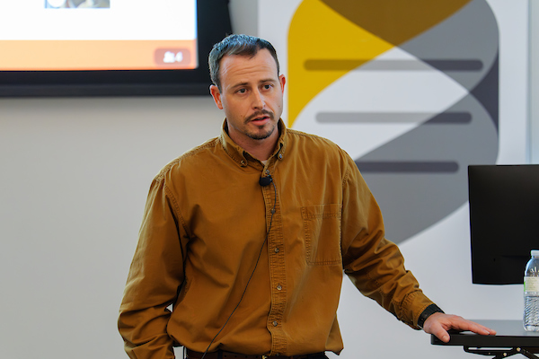
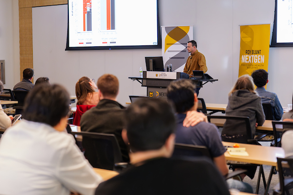

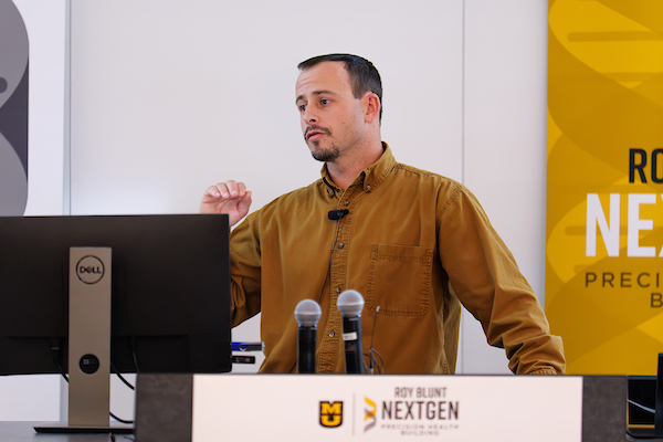
Dr. Zidao Wang’s Seminar 12/20/23
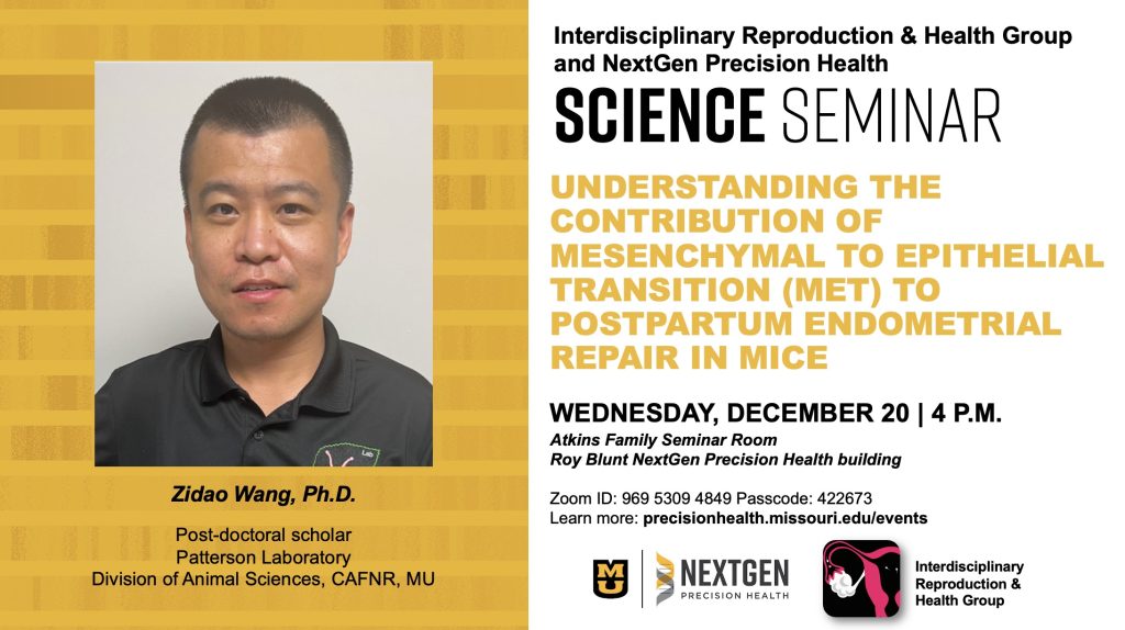
Understanding the contribution of mesenchymal to epithelial transition (MET) to postpartum endometrial repair in mice
Going through changes: understanding cell transition in the uterus after giving birth (lay audience summary of the seminar below)
Before we begin, what is mesenchymal-to-epithelial transition (MET)? This is a transition that happens during the generation of the innermost layer of the uterus, where mesenchymal cells change their identity to epithelial cells. During this transition, these cells lose their ability to move and become stationary. This process occurs during menstruation and after giving birth. Both MET and epithelial-to-mesenchymal transition (EMT) are important in processes such as organ formation, epithelial cell population management, and embryo development, but they are also involved in harmful processes such as the development of endometriosis and endometrial cancer.
Though MET is an important process that contributes to both healthy and disease states, it is poorly understood. Needing an appropriate mouse model, Dr. Zidao Wang, a post-doctoral scholar in the Division of Animal Sciences, University of Missouri, identified a type of mice that allowed him to efficiently “keep-tap” of mesenchymal cell identity throughout lifespan using the fluorescent signal. Using this mouse model, Dr. Wang investigated the progression of MET throughout the estrous cycle (mouse menstrual cycle) and found that there was no difference in populations of mesenchymal cells between estrus stages. When looking at mice throughout pregnancy and after birth, Dr. Wang found that there was an increase in MET after delivery with a peak at post-partum day 2 and mesenchymal cell populations returning to baseline by day 28 after giving birth.
In the future, he plans to investigate the mechanisms by which MET is triggered, how the epithelial cells are eventually replaced throughout reproduction, and what happens in situations where MET is dysregulated. The current and future work of Dr. Zidao will help the scientific community to better understand the mechanisms of important processes such as MET. (Written by Erin Carulli)
Dr. Lynda Harris’ Seminar 12/6/23


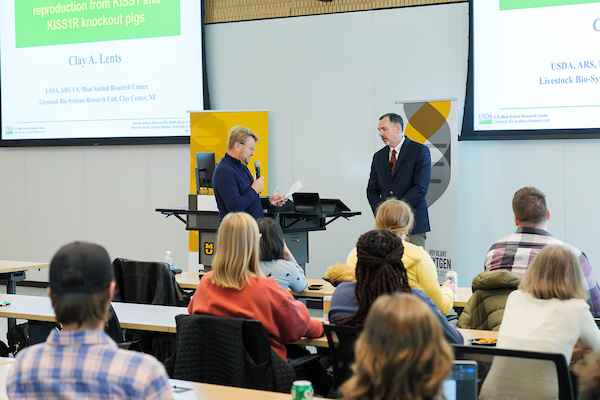
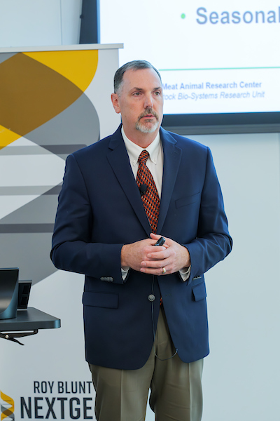
Dr. Clay Lents’ Seminar 11/1/23


Dr. Vincent Lynch’s Seminar 10/04/23


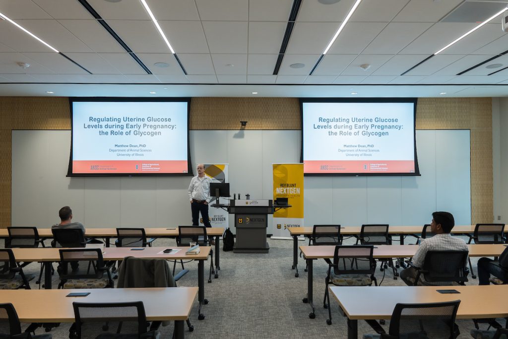
Dr. Matthew Dean’s Seminar 9/6/23


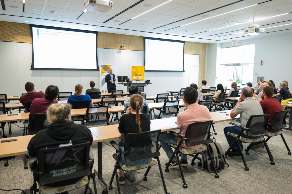
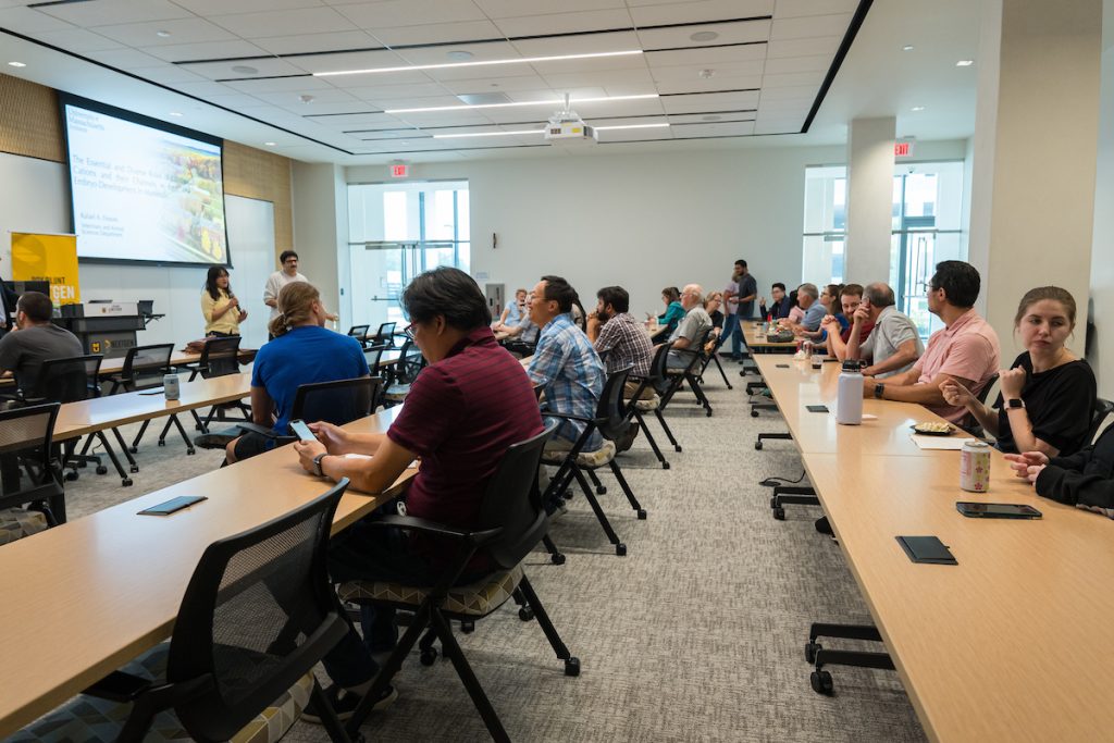
Dr. Rafael Fissore’s Seminar 8/2/23


Dr. Aileen Keating’s Seminar 5/3/23


IRHG outing event at International TAP House, 02/10/2023